Meningioma
What is a meningioma
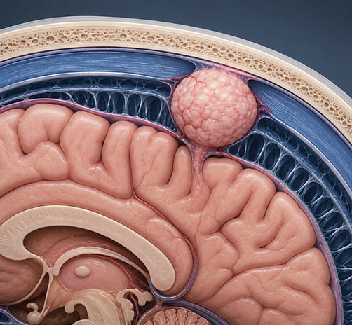
Meningioma is the most common primary brain tumour, accounting for approximately 20 to 30% of all intracranial tumours.
It arises from the meninges, the membranes that surround and protect the brain and spinal cord.
In the vast majority of cases, it is a benign tumour that grows slowly, compressing the surrounding brain tissue rather than infiltrating it, which often allows complete surgical removal.
Classification
Meningiomas are divided into three grades.
Grade I accounts for 85 to 90% of cases, is benign, grows slowly and usually remains stable for a long period of time.
Grade II, also known as atypical meningioma, occurs in approximately 20 to 30% of cases and has a greater tendency to recur.
Grade III, malignant meningioma, is rare, occurring in fewer than 2% of cases, grows more rapidly and is associated with a higher risk of recurrence after treatment.
Risk factors and epidemiology
Meningioma occurs about twice as often in women as in men, with incidence increasing particularly between the ages of 40 and 60, although it may also occur in younger or older patients.
It is associated with previous radiotherapy to the head, excessive exposure to X-rays, female hormones, as accelerated growth is sometimes observed during pregnancy, and hereditary syndromes such as neurofibromatosis type II.
In most cases, however, the exact cause remains unknown.
Approximately 30% of meningiomas are detected incidentally on imaging carried out for another reason, such as after head injury, while the simultaneous presence of more than one meningioma affects fewer than 10% of patients.
Symptoms
Many meningiomas remain completely asymptomatic for years, particularly when they are small. As the tumour grows, symptoms depend largely on its location, such as the convexity of the brain, the skull base or, more rarely, the spinal cord.
Headache that gradually worsens is common, while epileptic seizures, nausea, vomiting, visual disturbances such as diplopia or blurred vision, weakness in a limb, hearing loss, speech or balance disturbance, and changes in behaviour, memory or cognitive function may also occur.
When a meningioma is located in the spine, it may cause back pain or neurological deficits in the lower limbs.
Diagnosis
Diagnosis always begins with a detailed history and neurological examination, followed by imaging.
MRI with contrast is the investigation of choice, as it accurately demonstrates the location and size of the tumour and its relationship to surrounding structures, while CT helps to assess any bony involvement.
Definitive confirmation of histological grade is obtained through biopsy during surgery.

Treatment
Treatment of meningioma is not the same for every patient. It depends on the size and location of the tumour, the symptoms, the patient’s age and general health.
Observation
If the meningioma is small and not causing symptoms, the best option is often simple observation, with an MRI scan every 6 to 12 months. Many meningiomas never grow enough to require intervention.
Surgical resection
When the tumour causes symptoms or enlarges, surgery is the first option. The aim is to remove it as completely as possible, which significantly reduces the chance of it returning in the future. How easy or difficult that removal is depends mainly on the tumour’s location.
A meningioma on the surface of the brain is usually easier to access. A meningioma at the skull base, close to nerves and blood vessels, requires more specialised surgical technique in order to be removed safely.
Radiotherapy
In some cases, after surgery, or when the tumour’s location does not allow complete removal, additional radiotherapy is recommended. Modern techniques allow highly targeted delivery of radiation with great precision, limiting the effect on the healthy tissue around the tumour. Chemotherapy is used very rarely, only in very specific, treatment-resistant cases.
Dr Nektarios Mazarakis assesses each case individually in order to recommend the option best suited to each patient, whether that is simple observation, surgery or radiotherapy.
Prognosis
The prognosis for benign meningiomas is excellent, with five-year survival exceeding 90%. However, because of the possibility of recurrence even decades after the initial treatment, long-term imaging follow-up is required, particularly in cases of atypical or malignant meningiomas, where the risk of recurrence is clearly higher.
Book an Appointment
Dr Nektarios Mazarakis sees patients with meningiomas at his clinics in Athens and Thessaloniki, offering full clinical and imaging assessment in order to determine the most appropriate treatment approach.