Low-Grade Brain Tumours (Grade I-II)
What are low-grade brain tumours
A significant proportion of brain tumours fall into the low-grade category, also known as Grade I and Grade II tumours according to the World Health Organization classification (WHO CNS5, 2021 edition).
These tumours usually grow slowly and have a more favourable biological behaviour compared with high-grade tumours, although this does not mean that they do not require careful diagnosis, regular follow-up and, often, surgical treatment.
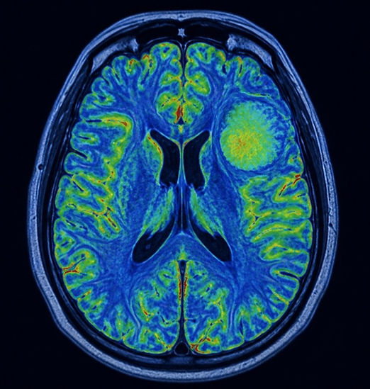
The modern classification of these tumours is no longer based solely on their histological appearance under the microscope, but also on molecular markers, such as mutation of the IDH gene and co-deletion of chromosomal regions 1p and 19q.
These markers now help to provide a more accurate prognosis in each case and to individualise the treatment approach.
Types of low-grade tumours
This category includes several different tumour types, depending on the tissue of origin.
Of glial, glioneuronal and ependymal origin
Pilocytic astrocytoma (Grade I): the most common low-grade tumour in children and young adults. It usually develops in the cerebellum, optic nerve or brainstem and has an excellent prognosis when completely removed surgically.
Subependymal giant cell astrocytoma, SEGA (Grade I): a rare tumour seen mainly in patients with tuberous sclerosis, usually near the foramen of Monro, with a possible risk of obstruction to cerebrospinal fluid flow.
Diffuse astrocytoma, IDH-mutant (Grade II): grows more diffusely within healthy brain tissue, without clear borders, which makes surgical removal more demanding.
Pleomorphic xanthoastrocytoma, PXA (Grade II): usually found in younger patients, in the temporal lobe, and often associated with a history of epileptic seizures.
Oligodendroglioma, IDH-mutant and 1p/19q co-deleted (Grade II): has a better response to treatment than purely astrocytic gliomas and tends to occur in the frontal lobes.
Ganglioglioma (Grade I): a mixed neuronal and glial tumour, a common cause of drug-resistant epilepsy in children and young adults.
Gangliocytoma (Grade I): a benign tumour composed exclusively of mature neuronal cells, with an excellent prognosis after surgical removal.
Dysembryoplastic neuroepithelial tumour, DNET (Grade I): often occurs in the temporal lobe in children and adolescents with a history of epilepsy, with a very good prognosis.
Central neurocytoma (Grade II): develops within the lateral ventricles of the brain and may cause hydrocephalus due to obstruction of cerebrospinal fluid flow.
Subependymoma (Grade I): usually an asymptomatic tumour discovered incidentally on imaging, with very slow progression.
Myxopapillary ependymoma (Grade I to II): found mainly in the lower part of the spinal cord, in the region of the conus medullaris and cauda equina.
Other tumour types in the same category
The broader category of low-grade brain tumours also includes meningioma, pituitary adenoma and craniopharyngioma.
Because of their different histological origin and distinctive clinical presentation, each of these tumour types is presented in detail on a separate page.
Read more about meningioma.
Read more about pituitary adenoma.
Read more about craniopharyngioma.
Symptoms
The symptoms of a low-grade brain tumour depend largely on its size, location and rate of growth.
Epileptic seizures are often the first symptom, particularly in diffuse gliomas occurring in younger adults. Headache, especially when persistent or gradually worsening, may also be a warning sign.
Depending on the location of the tumour, neurological deficits may occur, such as weakness in a limb, speech disturbance or visual disturbances.
In some cases, changes in cognitive function or behaviour are observed, particularly when the tumour affects the frontal lobes.
Larger tumours, or tumours causing hydrocephalus, may lead to signs of raised intracranial pressure, such as nausea, vomiting and impaired consciousness.
Diagnosis
MRI of the brain, with and without contrast, is the main investigation for detecting and initially characterising a low-grade tumour.
In many cases, additional functional MRI and tractography are performed in order to map critical functional areas of the brain before any surgical intervention.
The definitive diagnosis is established through histological and molecular examination of the tissue, either from a biopsy or from the specimen obtained during surgical resection. Testing for mutations such as IDH, as well as for 1p/19q co-deletion, is now essential for accurate tumour classification according to World Health Organization criteria.
Surgical management
Dr Nektarios Mazarakis, a neurosurgeon with subspecialist training in neuro-oncology through a national fellowship programme at Leeds General Infirmary in the United Kingdom, applies modern techniques of maximal safe resection for low-grade brain tumours. The aim of surgery is to remove as much of the tumour as possible without causing a new neurological deficit, especially when the tumour lies close to functionally critical areas of the brain.
In selected cases, intraoperative mapping or awake surgery is used in order to preserve functions such as speech and movement during tumour removal. The optimal surgical approach is always decided on an individual basis, within the framework of multidisciplinary collaboration with neuroradiologists and neuropathologists.
Prognosis and follow-up
The prognosis of low-grade tumours is generally more favourable than that of high-grade tumours. However, regular imaging follow-up is required, as some types, particularly diffuse astrocytomas and Grade II oligodendrogliomas, may over time progress to a higher grade of malignancy.
The molecular identity of the tumour is now the single most important prognostic factor.
Book an Appointment
Dr Nektarios Mazarakis sees patients with low-grade brain tumours at his clinics in Athens and Thessaloniki, offering individualised assessment based on his international experience in recognised neuro-oncology centres in the United Kingdom.